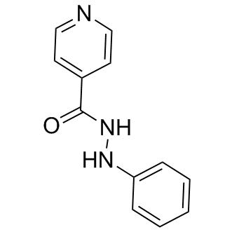
Additionally, the HL-60 leukemic cell line was used to assess whether TOP2 catalytic inhibitors can affect ANT LY2835219 CDK inhibitor cardiotoxicity without compromising their antiproliferative efficacy against leukemic cancer cells. The abilities of DEX, SOB and MER to chelate labile GDC-0879 intracellular Fe ions were assessed fluorimetrically using the calcein-AM assay. In H9c2 cells, a typical result was obtained with experimental Fe chelator SIH, which increased the intracellularly-trapped calcein fluorescence significantly compared to control from the start of incubation. DEX also increased significantly the calcein fluorescence compared to control, but only to,30% of the levels achieved with SIH. Moreover, a substantial lag-time was noticed as significant change in fluorescence compared to control was established only after,2.5 hours. Neither SOB nor MER were able to significantly displace Fe from its calcein-complex compared to control. During the last decade, both the traditional “ROS and Fe” hypothesis of ANT-induced cardiotoxicity as well as the notion that DEX protects cardiomyocytes via its Fe-chelating metabolite ADR-925 have been subjects of renewed discussion and growing controversy. Classical antioxidants and ROS scavengers, although usually very effective in acute ANT cardiotoxicity experiments, have failed in all controlled and randomised clinical trials conducted so far, although there were often notable discrepancies in the concentrations/doses of antioxidants used in in vitro and in vivo experiments as well as differences in the responses within various animal species. Furthermore, several Fe chelators that are both stronger and more selective for Fe than ADR-925 have failed to match the unique and experimental model-independent cardioprotective efficacy of DEX. Of significance, ADR-925 complexed with Fe may promote redox reactions rather than inhibiting them, and ADR-925 failed to protect isolated cardiomyocytes. In a model of chronic DAUinduced heart failure in rabbits, DEX was able to prevent increased cardiomyocyte apoptosis in hearts, but this protection was not accompanied by a significant reduction in lipoperoxidation, suggesting that oxidative stress may merely be a secondary by-product that accompanies ANT cardiotoxicity and subsequent heart failure rather than a primary culprit. Furthermore, a close derivative of DEX that lacks TOP2 inhibitory action but can be activated to produce a diacid diamide metabolite with ability to displace Fe from the complex with DOX has been found to be ineffective against chronic ANT cardiotoxicity in rats when compared to DEX. Thus, there has been a search for alternative or additional hypotheses to explain the mechanisms of both ANT cardiotoxicity as well as DEX-induced cardioprotection. The effective cardioprotective properties of DEX have been demonstrated by many clinical studies and were firmly confirmed with a recent meta-analysis. However, focused in vitro analyses of DEX cardioprotection and the mechanism involved have been scarce. Hasinoff et al. found that preincubation with DEX protected cardiomyocytes from mitochondrial membrane potential loss induced by relatively low concentrations of DOX. In the same study, the authors showed that signs of apoptosis are increased by approximately 200% after 48 h treatment with clinically relevant concentrations of DOX, which is comparable to our results obtained under similar conditions. In the present study, we employed a wide range of concentrations of DEX as well as two different ANTs to describe in detail the effects of these drugs at concentrations that can be present in the plasma of patients after drug administration. Furthermore, we directly compared these results in a model of oxidative insult induced by H2O2, which resulted in viability loss comparable  to both ANTs. Although DEX was able to consistently reduce LDH release from cardiomyocytes induced by DAU or DOX, it demonstrated no protection against any concentration of H2O2 employed in this study. Furthermore, the protective effects of DEX against DAU-induced toxicity were not associated with changes to GSH or GSSG within cardiomyocytes, which suggests that these effects are oxidative stress-independent. These results agree well with Lyu et al., who demonstrated that 200 mM DEX protected the H9c2 cardiomyoblast-derived cell line from DOX, but not H2O2- or camptothecin-induced DNA double strand breaks as determined by elevated c-H2AX expression.