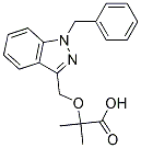
None of these compounds demonstrated selective cytotoxicity. In fact, most of these compounds were inactive. Together with their antitubulin activity, the selectivity of our compounds toward highly tumorigenic cell lines suggests that microtubules of tumorigenic and non-tumorigenic cell lines may differ. Interestingly, no difference in tubulin gene expression level was observed between highly tumorigenic and non-tumorigenic cell lines. It is plausible that observed selective cytotoxicity is not due to difference in tubulin gene expression but rather a result of differences in post-translational modifications. Recently, various experimental results have supported the notion that tubulin PTMs lead to the functional diversity of microtubules. Many tubulin PTMs have been identified including detrysosination, glutamylation, glycylation, acetylation phosphorylation and palmitoylation. Differences in tubulin isotype expression and PTMs have been associated with cell differentiation and developmental transitions. Because microtubules are key to mitotic spindle assembly and cell division, differences in mitotic spindle structure and function between tumorigenic and nontumorigenic cell lines may be associated with the selectivity of these compounds. In conclusion, we have identified a family of microtubule inhibitors that are mostly toxic against tumorigenic cell lines. Established cancer cell lines demonstrating high tumorigenicity in xenograft models may capture some properties of cancer cell subpopulations that are responsible for initiating and spreading the tumors. Therefore, we propose that this family of microtubule inhibitors, or related compounds with similar selectivity characteristics, BIBW2992 439081-18-2 should be considered as prime candidates for further evaluation as anticancer agents. Topo IIa inhibitors such as anthracyclines or epididophyllotoxins are important agents in the treatment of human SB431542 malignancy. These agents cause DNA damage by two mechanisms, locking Topo IIa in a cleavage complex producing DNA doublestrand breaks, and inhibiting chromatid decatenation. While the former mechanism is well understood, far less is known about the latter, yet it can be just as catastrophic to the cell. Failure of decatenation results in DSBs at anaphase, and to prevent this cells probably monitor decatenation at two positions in the cell cycle, at the G2/M boundary and at the metaphase to anaphase transition. These decatentation checkpoints are activated independently of the G2/M DNA damage-dependent checkpoint. Interestingly, lung and bladder cancers proceed through the decatenation checkpoints even in the presence of high levels of Topo IIa inhibitors, and this was thought to be secondary to a failure of the cell cycle arrest machinery. We recently isolated and characterized a human protein with SET and transposase domains called Metnase. Metnase promotes non-homologous end joining DNA repair, enhances plasmid and viral DNA integration, and cleaves but does not degrade supercoiled plasmid DNA. We recently showed that Metnase interacts with Topo IIa and enhances its function in chromosomal decatenation. Therefore, we hypothesized that Metnase may mediate the resistance of malignant cells to Topo IIa inhibitors, and chose to test this in breast cancer cells because anthracyclines are among the most important agents in the treatment of this disease. We report here that Metnase interacts with Topo IIa in breast cancer cells, promotes progression through metaphase in breast cancer cells treated  with a Topo IIa inhibitor, sensitizes breast cancer cells to the anthracycline adriamycin and the epididophyllotoxin VP16, and directly blocks Topo IIa inhibition by adriamycin in vitro. These data indicate that Metnase levels may be one reason why some breast cancer cells treated with Topo IIa inhibitors can progress through mitosis without catastrophe resulting in drug resistance. They have both an elevated Topo IIa level and significant Metnase expression.